色の認知には異なる光感受性物質を有する少なくとも2種類の網膜の光受容細胞を必要とする。人眼は最大の光感受性の光波長によって分類される3種類の錐体細胞を有している。それらは長波長感受性錐体(L-錐体;【旧】赤錐体)、中波長感受性錐体(M-錐体;【旧】緑錐体)、短波長感受性錐体(S-錐体;【旧】青錐体)であり、それぞれの最大光感受性波長はほぼ565, 545, 440nmである。これらの錐体細胞は外界からの光を生体電気信号に変換し、視中枢に伝達し色覚を誘発する。したがって、錐体光感受性細胞の欠損または不全は色覚異常を生じる。
先天色覚異常は1色覚、2色覚および異常3色覚の3種類のタイプに分類される。1色覚のほとんどは錐体細胞がすべて欠損し、網膜桿体細胞のみによる視覚を特徴とする。1色覚者はまた視力不良(低視力)、羞明(光に対する眩しさ)および眼球振盪を有する。1色覚は常染色体劣性遺伝形式をとる稀な症例であり、その頻度は0.003%とされる。2色覚と異常3色覚は以下の3種類に分類される:1型(L-錐体の不全または欠損)、2型(M-錐体の不全または欠損)および3型(S-錐体の不全または欠損)。これらのうち3型は常染色体優性遺伝形式をとり、先天青黄色覚異常を特徴とするが、その頻度は1色覚よりも低い。
先天色覚異常で最も多いタイプは赤緑異常を特徴とする1型色覚と2型色覚であり、これらはX染色体連鎖劣性遺伝形式をとり、その頻度は男性では4.5(日本)から10(欧米)%、女性では0.2(日本)から0.4(欧米)%である。
赤緑色覚異常者は正常色覚者よりも認識可能な色相が少ない。全ての色光のスペクトル(波長)は国際照明委員会(Commission Internationale de l'Ēclairage (International Commission onIllumination): CIE)による色度図上のスペクトル軌跡の各点に対応している。このスペクトル軌跡は馬蹄形としてあらわされ、その両端を結ぶ線分は紫系の勾配軌跡と呼ばれる。2色覚者では3原色の替わりに2原色を基にスペクトル色に対応している。赤緑2色覚者は540 nm以上の波長の光に対して、ほぼ540 nmのスペクトル光で対応している。2色覚者は490 nmと500 nmの間の波長の相違を識別する能力(波長識別能)が良好であるが、この波長域の両側では識別能が急激に低下する。2色覚者が識別不能である色はCIE色度図上の同一軌跡上に直線状に分布し、混同色軌跡と呼ばれる。正常色覚者は波長495 nmと500 nmの光を青緑として認識するが、1型色覚者および2型色覚者はそれぞれ495 nmと500 nmのスペクトル光を等エネルギー光である白色光と混同する。これらのスペクトル光点は中性点または無彩色点と呼ばれる。この混同色軌跡は1型色覚者と2型色覚者とで異なる。加えて、異常3色覚者は正常色覚者と2色覚者との間に位置し,色覚の個人差が大きい。また、2型色覚者および正常色覚者と比較して1型色覚者は長波長光に対する感受性が低いことが特徴として挙げられる。したがって、1型色覚者は正常3色覚者よりも赤色をより暗く感じる。
色覚異常者は正常3色覚者が認識できる色に関してその認識・識別に困難さが生じる。特に、2色覚者は色を正常色覚者とは異なる色として認識(誤認)することが多く,時には学校生活や職業上,多大な影響が生じる場合がある。程度の差はあるが,異常3色覚者は2色覚者よりも色の認識での制約は軽度であるが、条件によっては色の誤認が起きる場合があり得る。先天色覚異常者は自己の色覚と正常色覚者の色覚との差を自覚することが困難である可能性が高い。色覚は社会的独自性の上で修得、形成される一方で、様々な実生活面で重要な意義を有している。したがって、個人と実生活との共生のために早い時期において色覚異常の有無に関して正しい診断を受けておくことが重要である。
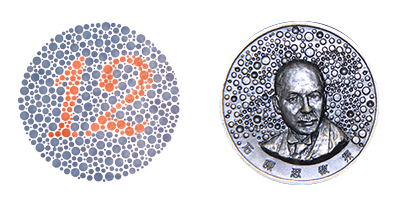
1933年、第14回国際眼科学会(マドリード、スペイン)において石原色覚検査表、スチリング(Stilling)色覚検査表およびナーゲル(Nagel)アノマロスコープが色覚異常の標準検査法として推奨された。特に、石原色覚検査表は臨床その他の場所で容易に使用でき、かつ他の同様な仮性同色表と比較して、検査としての特異度(正常色覚を色覚異常と判定する偽陽性度が低い)と感度(色覚異常を正常色覚として判定する偽陰性度が低い)とがともに高いことが標準的検査表として推奨された最大の根拠である。
石原色覚検査表は1916年に東京大学医学部眼科学講座 石原 忍教授によって考案された。当初は石原色盲検査表と命名され、16表で構成されていた。その後、改良ならびに表数が増加され、1936年に32表、1951年に38表となった。
公益財団法人一新会は色覚検査表の品質の維持および改良ならびに色覚異常の検査表に関する研究支援を目的に石原色覚検査表の版権を基に1961年に財団法人として設立された。大熊篤二をはじめとする多くの卓越した研究者が財団法人の目的に従っての様々な色覚の研究に参加、尽力した。1980年に大熊教授は独自の仮性同色表を創作し、新色覚異常検査表として発表した。この検査表の色覚異常の検査に関する特異度と感度は石原色覚検査表と同様の成績が得られている。今回、財団法人一新会は平成24年度に公益財団法人としての再発足することもあり、石原色覚検査表および新色覚異常検査表の中から検査能力に特に優れた検査表を基に新たな石原色覚検査表IIを編纂、上梓することとした。
公益財団法人 一新会
Preface
The generation of colour perception requires at least two types of retinal photoreceptor cells containing different light-sensitive pigments. Human eyes contain 3 types of cone photoreceptor cells that are classified by their different wavelengths of maximum light sensitivity. They comprise long wavelength sensitive cones (L-cones), middle wavelength sensitive cones (M-cones) and short wavelength sensitive cones (S-cones) and their wavelengths of maximum light sensitivity are approximately 565, 545 and 440nm, respectively. These cone cells mediate the conversion of light into electrical signals that evoke colour perception. Therefore, defective or missing cone photoreceptor cells cause defective colour vision.
Congenital colour vision deficiency can be classified into three types; monochromatism, dichromatism and anomalous trichromatism. Most cases of monochromatism are characterized by an absence of cone cells, leaving rods alone to sense visual stimuli. Individuals with this condition also have poor visual acuity, light sensitivity (photophobia) and nystagmus. Monochromatism is a rare autosomal recessive disorder with an incidence of 0.003%. Dichromatism and anomalous trichromatism are classified into three types; protan (anomalous or missing L-cones), deutan (anomalous or missing M-cones) and tritan (anomalous or missing S-cones). Tritan defects are caused by an autosomal dominant condition. They are characterized by congenital blue-yellow deficiency and have an incidence less than that of monochromatism.
The most frequent types of congenital colour vision deficiency are protan and deutan, which are red-green colour deficiencies. These X-linked recessive disorders have an incidence of 4.5-10% in men and 0.2-0.4% in women.
Individuals with red-green deficiency can see fewer colours than those with normal colour vision. All colors of spectral, or single-wavelength, lights correspond to points on the spectrum locus of the CIE (Commission Internationale de l'Ēclairage, International Commission onIllumination ) chromaticity diagram. This is an open, horseshoe-shaped curve with the two ends connected by a line representing the gradient of purple colors. Dichromats need two primary colors instead of three primary colors to match spectrum colors. Red-green dichromats can nearly match a 540 nm spectral light to all wavelengths above 540 nm. Dichromats are able to discriminate differences in wavelength between 490 and 500 nm, but discrimination deteriorates markedly outside of this range. Colours that the dichromat cannot discriminate are located on straight lines in the CIE chromaticity diagram, i.e., isochromatic lines. Subjects with normal colour vision perceive light with wavelengths of 495 and 500nm as blue-green. Protanopes and deuteranopes confuse equal-energy white lights with 495 nm and 500 nm spectral lights, respectively. These spectral points are called neutral, or achromatic, points. Additionally, there are large phenotypical variations in the colour vision of individuals with dichromatic and anomalous trichromatic deficiencies. It is noteworthy that both protanopes and protanomals are less sensitive to long wavelengths than deutans and those with normal colour vision. Hence, protanopes perceive red as being darker than it appears to normal trichromats.
Colour vision deficiencies lead to difficulties in identifying the full range of colours seen by normal trichromats. In particular, dichromats may fail to see or discriminate colours needed to properly perform tasks in both school and work life. The anomalous trichromat has less restriction than the dichromat, but may have trouble with colour perception in particular situations. Individuals with congenital colour deficiency may not be aware that their color perception differs from those with normal colour vision. Color vision is an integral part of social independence, impacting many aspects of life. Hence, it is imperative that the diagnosis of color deficiency be made early in life.
In 1933, Ishihara’s Test, Stilling’s Test and Nagel’s Anomaloscope were recognized at the 14th World Ophthalmology Congress in Madrid, Spain as standard test methods to measure color deficiencies. In particular, Ishihara’s Test is portable and easily administered with high specificity (low false positives) and high sensitivity (low false negatives) when compared to other pseudo isochromatic test plates.
Ishihara’s Test for Colour Deficiency was invented by Professor Shinobu Ishihara at the University of Tokyo, Japan in 1916. It was originally named Ishihara’s Test for Colour Blindness and was composed of 16 plates. The set of test plates was increased to 32 plates in 1936 and 38 plates in 1951. The Isshinkai Foundation was founded in 1961 based on the copyright of the Ishihara’s Test to qualify the Test and to support the investigation of tests for colour deficiency. Many distinguished researchers such as Professor Tokuji Okuma endeavored to study various aspects of colour vision according to the mission of the Foundation. In 1980, Professor Okuma developed his own pseudo isochromatic test plates called the New Test for Colour Deficiency. The sensitivity and specificity of this test in the detection of color deficiency is similar to that of Ishihara’s Test. Therefore, the Foundation published the Ishihara’s Test for Colour Deficiency II which included both Ishihara’s and Okuma’s test plates. The Ishihara’s Test for Colour Deficiency II has high sensitivity and specificity to detect anomalous colour vision.
Public Interest Incorporated Foundation
ISSHINKAI
Tokyo, Japan
※「石原式」と「ISHIHARA TEST」は、登録商標です。
※ The trademarks (「石原式」and「ISHIHARA TEST」) are registered on the Japan Patent Office.